
Complete Resection of A Giant Convexity Meningioma Involving The Middle Cerebral Artery: A Case Report and Surgical Considerations
A Case Report
DOI:
https://doi.org/10.33005/jdiversemedres.v3i7.371Keywords:
Convexity meningioma, middle cerebral artery, simpson grading, surgical resectionAbstract
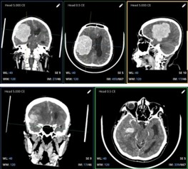
Meningioma is the most common primary intracranial tumor in adults, accounting for approximately 37.6% of all primary central nervous system neoplasms. Although convexity meningiomas are generally surgically accessible, large tumors with neurovascular involvement present significant operative challenges, especially when acute neurological deterioration necessitates emergent intervention. A 46-year-old female presented with decreased consciousness (GCS E2V3M4) and progressive hemiparesis. Preoperative CT revealed a large temporoparietal extra-axial mass with significant mass effect and middle cerebral artery (MCA) involvement. Emergent craniotomy and stepwise debulking were performed utilizing gravity-assisted retraction, central dural incision, pial plane preservation, early arterial feeder coagulation, and systematic hemostasis. The philosophy of prioritizing patient safety over gross total resection guided the strategy. Postoperatively, the patient regained full consciousness (GCS E4V5M6) with marked motor improvement. Emerging evidence supports subtotal resection with adjuvant stereotactic radiosurgery for residual tumors. Emergent surgical debulking of large convexity meningiomas with neurovascular involvement is feasible and effective when guided by stepwise resection, pial plane preservation, and patient safety. Adjuvant radiosurgery should be considered for any residual tumor on follow-up imaging.
Downloads
References
Ogasawara C, Philbrick BD, Adamson DC. Meningioma: A review of epidemiology, pathology, diagnosis, treatment, and future directions. Biomedicines 2021;9(3):319; doi: 10.3390/biomedicines9030319.
Pacult MA, Przybylowski CJ, Raza SM, et al. Surgical Management of High-Grade Meningiomas. Cancers (Basel) 2024;16(11):1978; doi: 10.3390/cancers16111978.
Brokinkel B, Spille DC, Brokinkel C, et al. The Simpson grading: defining the optimal threshold for gross total resection in meningioma surgery. Neurosurg Rev 2021;44(3):1713–1720; doi: 10.1007/s10143-020-01369-1.
Gagnier JJ, Kienle G, Altman DG, et al. The CARE guidelines: Consensus-based clinical case reporting guideline development. J Med Case Rep 2013;7(1):223; doi: 10.1186/1752-1947-7-223.
SIMPSON D. The recurrence of intracranial meningiomas after surgical treatment. J Neurol Neurosurg Psychiatry 1957;20(1):22–39; doi: 10.1136/jnnp.20.1.22.
Voß KM, Spille DC, Sauerland C, et al. The Simpson grading in meningioma surgery: does the tumor location influence the prognostic value? J Neurooncol 2017;133(3):641–651; doi: 10.1007/s11060-017-2481-1.
Hasseleid BF, Meling TR, Rønning P, et al. Surgery for convexity meningioma: Simpson Grade I resection as the goal - Clinical article. J Neurosurg 2012;117(6):999–1006; doi: 10.3171/2012.9.JNS12294.
Otero-Rodriguez A, Tabernero MD, Munoz-Martin MC, et al. Re-Evaluating Simpson Grade I, II, and III Resections in Neurosurgical Treatment of World Health Organization Grade I Meningiomas. World Neurosurg 2016;96:483–488; doi: 10.1016/j.wneu.2016.09.007.
Teng H, Yu J, Pang Y, et al. Long-term recurrence risk and temporal dynamics after resection of intracranial meningiomas: A multicenter cohort study with up to 15 years of follow-up. Cancer 2025;131(23):e70200; doi: 10.1002/cncr.70200.
Beighley A, Kesbeh Y, Rahimian J, et al. Impact of Fractionation Regimen on Local Control Following Frameless Linear Accelerator-Based Image-Guided Stereotactic Radiosurgery and Radiotherapy for Intracranial Meningioma. World Neurosurg 2024;190:e403–e412; doi: 10.1016/j.wneu.2024.07.148.
Wang A, Zhang X, Sun KK, et al. Deep Sylvian fissure meningiomas: A case report. World J Clin Cases 2022;10(21):7438–7444; doi: 10.12998/wjcc.v10.i21.7438.
Goldbrunner R, Stavrinou P, Jenkinson MD, et al. EANO guideline on the diagnosis and management of meningiomas. Neuro Oncol 2021;23(11):1821–1834; doi: 10.1093/neuonc/noab150.
World Health Organization Grade I Convexity Meningiomas: Study on Outcomes, Complications and Recurrence Rates. [Internet]. 2015 [cited 2026 Aug 13].
Outcome After Surgical Resection Of Giant Intracranial Meningiomas: Clinical and Radiological study. DOAJ. 2026.
The Management of a Giant Convexity en Plaque Anaplastic Meningioma with Gerstmann Syndrome: A Case Report of Surgical Outcomes in a 76-Year-Old Male. PMC. 2024
World Health Organization. Central nervous system tumours. In: WHO Classification of Tumours. 5th ed. Lyon: IARC; 2021.
Downloads
Published
How to Cite
Issue
Section
License
Copyright (c) 2026 Journal of Diverse Medical Research : Medicosphere

This work is licensed under a Creative Commons Attribution 4.0 International License.
CC Attribution 4.0





 This journal applies the Creative Commons Attribution 4.0 International License (CC BY 4.0), which permits unrestricted use, distribution, and reproduction in any medium, provided the original author and source are properly credited.